 US & Canada:
  Email:


 Access IFU & SDS
Summary & Explanation
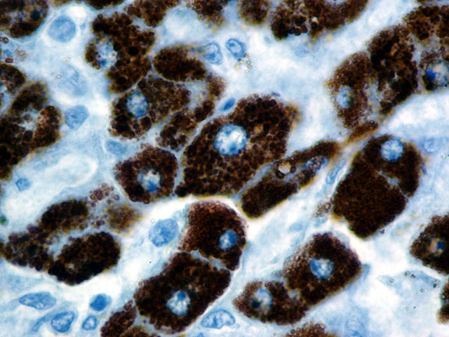
{"type":"root","children":[{"type":"paragraph","children":[{"type":"text","value":"Hepatocyte Specific Antigen (HSA or Hep Par 1) has been demonstrated consistently in the vast majority of Hepatocellular Carcinomas. Studies have shown the utility of HSA in the differential diagnosis of Hepatocellular Carcinoma, Cholangiocarcinoma and Hepatoblastomas."}]},{"type":"paragraph","children":[{"type":"text","value":"HSA recognizes both benign and malignant liver derived tissues including such tumors as Hepatoblastoma, Hepatocellular Carcinoma, and Hepatic Adenoma. It recognizes both normal adult and fetal liver tissue. The typical pattern is a granular cytoplasmic staining. This antibody is useful in differentiating Hepatocellular\n Carcinomas with adenoid features from Adenocarcinomas, either primary in the liver or metastatic lesions to the liver. In recognizing Hepatoblastoma, it is useful in differentiating this entity from other small round cell tumors."}]}]}
Antibody Type
Mouse Monoclonal
Clone
OCH1E5
Isotype
IgG1/K
Localization
Cytoplasmic
Dilution
1:100-1:500
Species Reactivity
Human, Dog, Cat
Immunogen
Formalin-fixed, failed human allograft liver that was mechanically disrupted.