 US & Canada:
  Email:


 Access IFU & SDS
Summary & Explanation
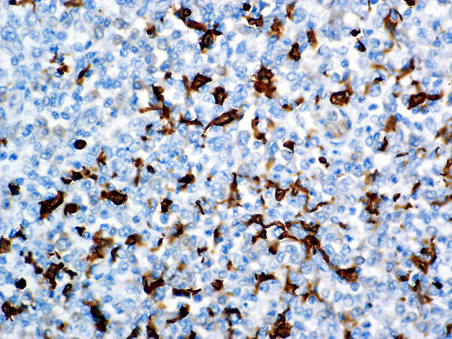
{"type":"root","children":[{"type":"paragraph","children":[{"type":"text","value":"Fascin, encoded by the human homolog for sn (hsn) gene, has been localized to microspikes and stress fibers of cultured cells where it is thought to be involved in the formation of microfilament bundles. It is expressed predominantly in dendritic cells. Lymphoid cells, myeloid cells and plasma cells are negative. However, Reed Sternberg cells in Hodgkin’s Lymphoma are positive for Fascin staining. Epstein-Barr virus may induce expression of Fascin in B-cells."}]},{"type":"paragraph","children":[{"type":"text","value":"Fascin is a very sensitive marker for Reed-Sternberg cells and variants in nodular sclerosis, mixed cellularity, and lymphocyte depletion Hodgkin’s Disease. This marker might be helpful in distinguishing between Hodgkin’s Disease and Non-Hodgkin Lymphoma in difficult cases. Also, the lack of expression of Fascin in the\n neoplastic follicles in Follicular Lymphoma can be helpful in distinguishing these lymphomas from reactive Follicular Hyperplasia in which the number of follicular dendritic cells is normal or increased."}]}]}
Antibody Type
Mouse Monoclonal
Clone
BSB-36
Isotype
IgG2a/K
Localization
Cytoplasmic
Dilution
1:250-1:1000
Species Reactivity
Human, Rat
Immunogen
Fascin purified from HeLa cells.