 US & Canada:
  Email:


 Access IFU & SDS
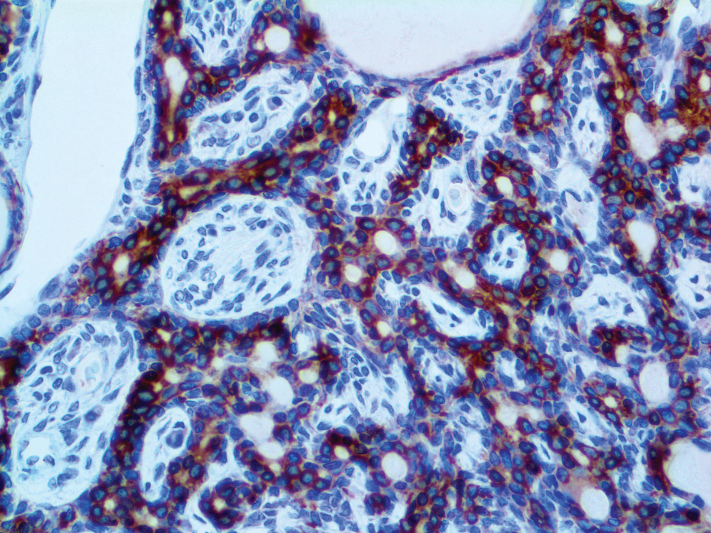
Summary & Explanation
{"type":"root","children":[{"type":"paragraph","children":[{"type":"text","value":"Cytokeratins are intermediate-filament keratins found in the intracytoplasmic cytoskeleton of epithelial tissue. There are two types of cytokeratins: the low-weight, acidic Type I cytokeratins and the high-weight, basic or neutral Type II cytokeratins. Cytokeratins are usually found in pairs comprising a Type I cytokeratin and a Type II cytokeratin. Expression of these cytokeratins is frequently organ or tissue-specific."}]},{"type":"paragraph","children":[{"type":"text","value":"Cytokeratin Low Molecular Weight AE1 can recognize most acidic keratins, making it a broadly reactive antibody that stains most epithelia and their neoplasms. Members of the acidic and basic subfamilies are found in pairs. Each epithelium contains at least one acidic and one basic keratin so this antibody can show the distribution of keratin-containing cells in epithelia. Cytokeratin AE1 is particularly suited to distinguish poorly-differentiated Carcinomas from non-epithelial Neoplasms. This marker stains both normal and neoplastic cells of epithelial origin."}]}]}
Antibody Type
Mouse Monoclonal
Clone
AE1
Isotype
IgG1
Localization
Cytoplasmic
Dilution
1:100-1:500
Species Reactivity
Human, Mouse, Rat, Monkey, Rabbit, Chicken, Cattle, Shrew, Fish
Immunogen
Purified human epidermal keratin.