 US & Canada:
  Email:


 Access IFU & SDS
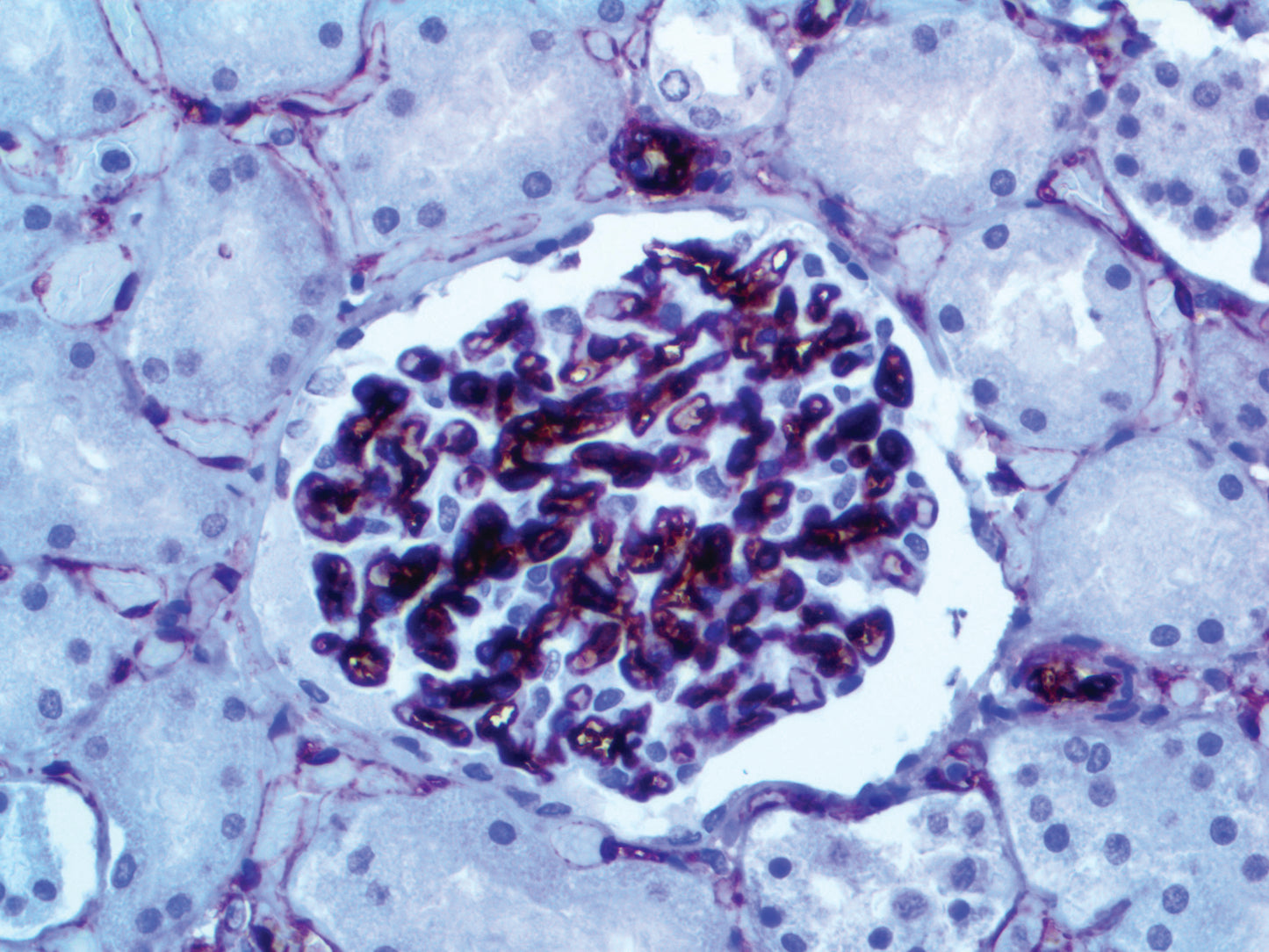
Summary & Explanation
{"type":"root","children":[{"type":"paragraph","children":[{"type":"text","value":"CD34 functions as a cell-cell adhesion factor and cell-surface glycoprotein. It may also mediate the attachment of stem cells to bone marrow extracellular matrixes or directly to stromal cells. Cells expressing CD34 are normally found in the umbilical cord and bone marrow as hematopoietic cells, and in vascular endothelium. In addition to stem cell recognition, CD34 is expressed by vascular endothelium; it appears that proliferating endothelial cells express this molecule in greater amounts than resting cells. In comparison to factor VIII R Antigen, CD34 stains are stronger and appear to be more sensitive in nature."}]},{"type":"paragraph","children":[{"type":"text","value":"In tumors, CD34 is found in Alveolar Soft Part Sarcoma, pre B-ALL (positive in 75%), AML(40%), AMLM7 (most), Dermatofibrosarcoma Protuberans, Gastrointestinal Stromal Tumors, Giant Cell Fibroblastoma, Granulocytic Sarcoma, Kaposi’s Sarcoma, Liposarcoma, Malignant Fibrous Histiocytoma, Malignant Peripheral Nerve Sheath tumors, Meningeal Hemangiopericytomas, Meningiomas, Neurofibromas, Schwannomas, and Papillary Thyroid Carcinoma. A negative CD34 may exclude Ewing’s Sarcoma/PNET, Myofibrosarcoma of the breast, and Inflammatory Myofibroblastic tumors of the stomach."}]}]}
Antibody Type
Mouse Monoclonal
Clone
QBEnd\10
Isotype
IgG1
Localization
Cytoplasmic, Membranous
Dilution
1:100-1:500
Species Reactivity
Human, Dog
Immunogen
Human endothelial vesicles.