 US & Canada:
  Email:


 Access IFU & SDS
Summary & Explanation
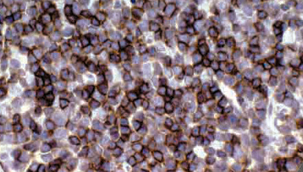
{"type":"root","children":[{"type":"paragraph","children":[{"type":"text","value":"CD1 proteins have been demonstrated to restrict T-cell response to non-peptide lipid and glycolipid antigens. At least five CD1 genes (CD1a, b, c, d, and e) have been identified. CD1a belongs to a family of glycoproteins expressed on the surface of various human antigen-presenting cells. In particular, CD1a is a protein of 43 to 49 kDa, and has been shown to be expressed on dendritic cells and cortical thymocytes. Langerhans cells in the skin and some epithelia also express this protein. This antigen is expressed in cells comprising Langerhans Cell Histiocytosis and Langerhans Cell Sarcoma."}]},{"type":"paragraph","children":[{"type":"text","value":"Anti-CD1a has been used to differentiate various cutaneous Lymphomas (T-cell) from B-cell Lymphomas and Pseudolymphomas. CD1a is also expressed by some malignancies of T-cell lineage and in Histiocytosis X."}]}]}
Antibody Type
Rabbit Monoclonal
Clone
EP80
Isotype
IgG
Localization
Cytoplasmic, Membranous
Dilution
1:50-1:200
Species Reactivity
Human
Immunogen
Synthetic peptide corresponding to residues in human CD1a protein.