 US & Canada:
  Email:


 Access IFU & SDS
Summary & Explanation
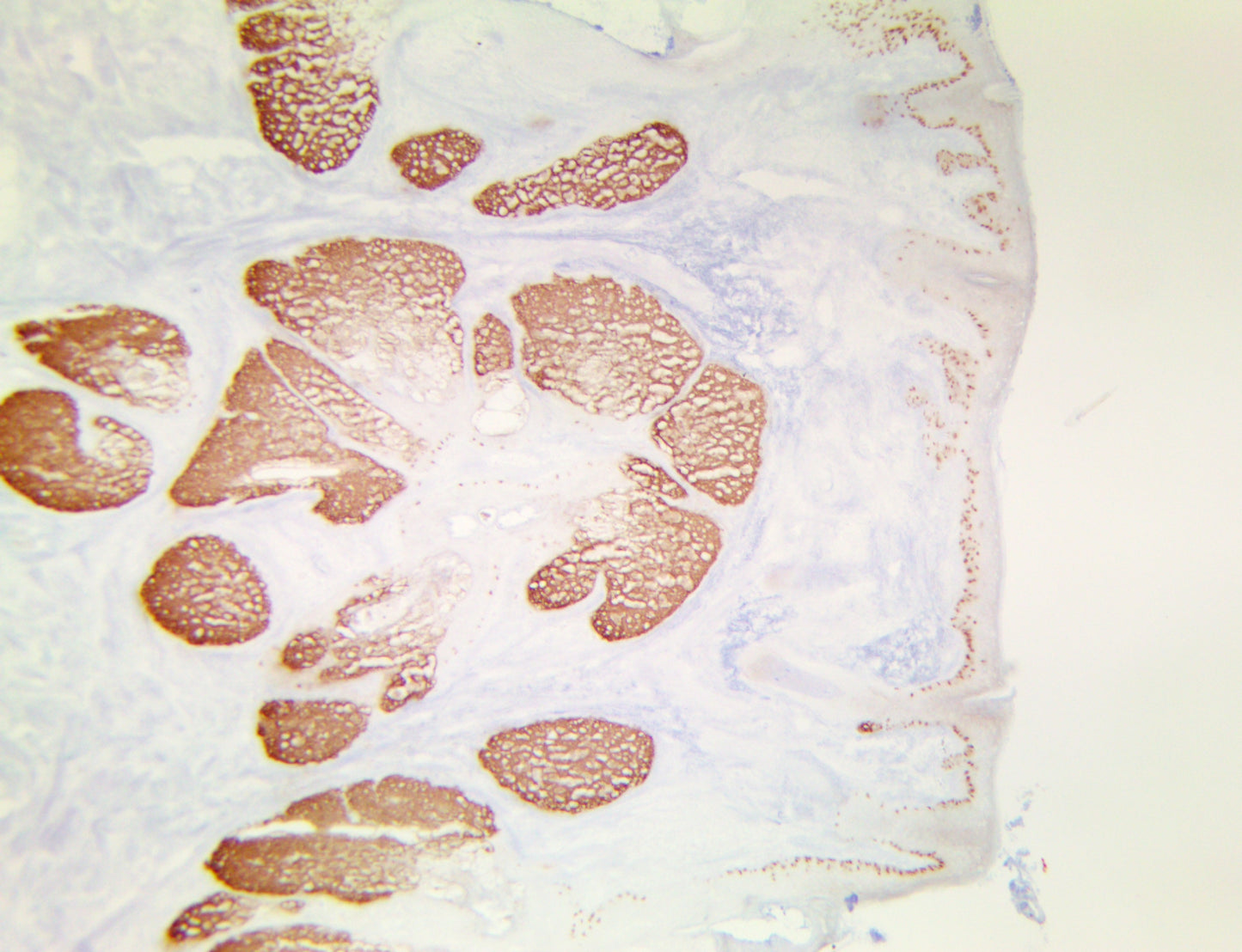
{"type":"root","children":[{"type":"paragraph","children":[{"type":"text","value":"Melanoma antigen preferentially expressed in tumors is a protein that in humans is encoded by the PRAME gene. This gene encodes an antigen that is predominantly expressed in human Melanomas and is recognized by cytolytic T lymphocytes. PRAME is not expressed in normal tissues, except in testis. This expression pattern is like that of other CT antigens, such as MAGE, BAGE and GAGE. However, unlike these other CT antigens, this antigen is also expressed in Acute Leukemias."}]},{"type":"paragraph","children":[{"type":"text","value":"PRAME overexpression in triple negative breast cancer has also been found to promote cancer cell motility through induction of the epithelial-to-mesenchymal transition. PRAME mRNA expression is well documented in Cutaneous and Ocular Melanomas. One study concluded that diffuse nuclear immunoreactivity for PRAME was found in 87% of metastatic and 83.2% of primary Melanomas. Among Melanoma subtypes, PRAME was diffusely expressed in 94.4% of acral Melanomas, 92.5% of superficial spreading Melanomas, 90% of Nodular Melanomas, 88.6% of Lentigo Maligna Melanomas, and 35% of Desmoplastic Melanomas. When in situ and NonDesmoplastic Invasive Melanoma components were present, PRAME expression was seen in both. Most Melanocytic nevi (86.4%), were completely negative for PRAME."}]},{"type":"paragraph","children":[{"type":"text","value":"Immunoreactivity for PRAME was seen, albeit usually only in a minor subpopulation of lesional Melanocytes, in 13.6% of Cutaneous Nevi, including Dysplastic Nevi, Common Acquired Nevi, Traumatized/recurrent Nevi, and Spitz Nevi. Rare isolated junctional Melanocytes with immunoreactivity for PRAME were also seen in Solar Lentigines and benign nonlesional skin. This study suggests that immunohistochemical analysis for PRAME expression may be useful for diagnostic purposes to support a suspected diagnosis of Melanoma. It may also be valuable for margin assessment of a known PRAME-positive Melanoma, but its expression in Nevi, Solar Lentigines, and Benign nonlesional Skin can represent a challenge."}]}]}
Antibody Type
Rabbit Monoclonal
Clone
RBT-PRAME
Isotype
IgG
Localization
Nuclear
Species Reactivity
Human
Immunogen
Synthetic peptide corresponding to residues of human PRAME protein.