 US & Canada:
  Email:


 Access IFU & SDS
Summary & Explanation
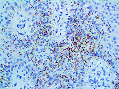
{"type":"root","children":[{"type":"paragraph","children":[{"type":"text","value":"Treponema pallidum is a spirochaete bacterium. The treponemes have a cytoplasmic and an outer membrane. The shape of T. pallidum is flat and wavy, unlike the other spirochetes, which are helical. Using light microscopy, treponemes are only visible using dark field illumination. They are Gram negative, but some regard them too thin to be Gram stained. T. pallidum is a motile spirochaete that is generally acquired by close sexual contact, entering the host via breaches in squamous or columnar epithelium. The organism can also be transmitted to a fetus by transplacental passage during the later stages of pregnancy, giving rise to congenital syphilis. The helical structure of T. pallidum allows it to move in a corkscrew motion through a viscous medium such as mucus. It gains access to the host’s blood and lymph systems through tissue and mucous membranes."}]},{"type":"paragraph","children":[{"type":"text","value":"Detection of Treponema pallidum can be difficult, and the correct diagnosis of secondary syphilis is critical. Diagnosis of syphilis is usually based on clinical presentation, dark-field microscope analysis, and serological tests. T. pallidum can be also evidenced by Immunohistochemistry in up to 90% of the samples with the bacteria located in the epidermis and the upper dermis of formalin-fixed paraffin-embedded tissues."}]}]}
Antibody Type
Rabbit Polyclonal
Clone
Polyclonal
Isotype
IgG
Localization
Cell Wall
Dilution
1:250-1:1000
Species Reactivity
Eubacteria
Immunogen
Purified treponema pallidum.