 US & Canada:
  Email:


 Access IFU & SDS
Summary & Explanation
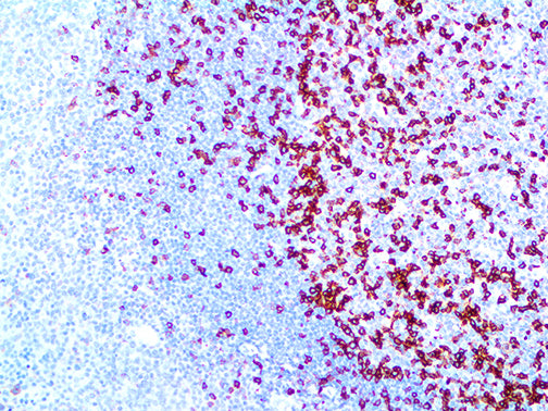
{"type":"root","children":[{"type":"paragraph","children":[{"type":"text","value":"Programmed Death 1 (PD-1 or CD279) is a Type I membrane protein comprised of 268 amino acids. PD-1 is a member of the extended CD28/CTLA-4 family of T-cell regulators. PD-1 is expressed on the surface of activated T-cells, B-cells, and macrophages. In comparison to CTLA-4, PD-1 more broadly negatively regulates immune responses. New data suggests that expression of PD-L1 on tumor cells inhibits anti-tumor activity through engagement of PD-1 on effector T-cells. Expression of PD-L1 on tumors is correlated with reduced survival in esophageal, pancreatic and other types of cancers, highlighting the relevance of exploring the PD-1 pathway as a target for immunotherapy. Studies have found that PD-1 is expressed on most T-cells and a small subset of B-cells in the light zone of germinal centers, but not elsewhere in the tonsil. On that basis, it was postulated that PD-1 may play a role in the process of clonal selection of centrocytes, which occurs in this subanatomic site in germinal centers. PD-1 is a new marker of Angioimmunoblastic Lymphoma and suggests a unique cell of origin for this neoplasm. Unlike CD10 and bcl-6, PD-1 is expressed by few B-cells, so it may be a more specific and useful diagnostic marker in Angioimmunoblastic Lymphoma. It also seems to stain a greater percentage of CD3-positive neoplastic cells in Angioimmunoblastic Lymphoma than either CD10 or bcl-6."}]}]}
Antibody Type
Rabbit Monoclonal
Clone
EP239
Isotype
IgG
Localization
Cytoplasmic, Membranous
Dilution
1:50-1:200
Species Reactivity
Human
Immunogen
A synthetic peptide corresponding to resides of human PD-1 protein.