 US & Canada:
  Email:


 Access IFU & SDS
Summary & Explanation
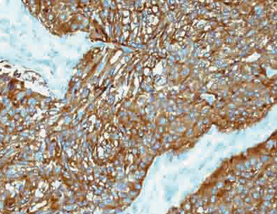
{"type":"root","children":[{"type":"paragraph","children":[{"type":"text","value":"Synaptophysin is a synaptic vesicle glycoprotein weighing 38 kDa. It is present in endocrine cells, the brain, spinal cord, and adrenal glands. It acts as a marker for neuroendocrine cells."}]},{"type":"paragraph","children":[{"type":"text","value":"Synaptophysin reacts with neuroendocrine cells of human adrenal medulla, carotid body, skin, pituitary, thyroid, lung, pancreas and gastrointestinal mucosa. Positive staining is seen in neurons of the brain, spinal cord, retina, and Paneth’s cells in the gastrointestinal tract and gastric parietal cells. This antibody identifies normal neuroendocrine cells and neuroendocrine neoplasms. Diffuse, finely-granular cytoplasmic staining is observed and probably correlates with the distribution of the antigen within neurosecretory vesicles. The expression of Synaptophysin is independent of the presence of NSE or other neuroendocrine markers. Synaptophysin is an independent broad-range marker of neural and neuroendocrine differentiation."}]}]}
Antibody Type
Rabbit Monoclonal
Clone
EP158
Isotype
IgG
Localization
Cytoplasmic
Dilution
1:100-1:500
Species Reactivity
Human, Predicted: Mouse, Rat, Donkey
Immunogen
A synthetic peptide corresponding to residues on the C-terminus (cytoplasmic domain) of human Synaptophysin protein.