 US & Canada:
  Email:


 Access IFU & SDS


Summary & Explanation
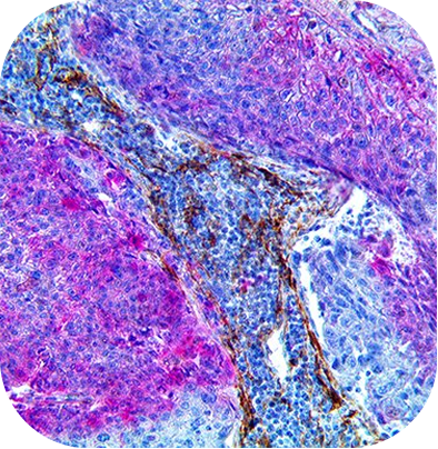
{"type":"root","children":[{"type":"paragraph","children":[{"type":"text","value":"In cutaneous squamous and basal cell carcinoma, Perineural Invasion (PNI) is the infiltration of tumor within the perineural space. PNI is an uncommon manifestation of SCC and BCC but can indicate adverse outcomes including recurrence, metastasis, poor prognosis, and death. During microscopically controlled excision, Immunohistochemistry (IHC) may increase accuracy in assessing the presence of PNI, especially in smaller nerves. IHC may be more accurate than routine sections in diagnosing PNI, with significant implications for patient staging, prognosis, and management. Multiplex IHC may further improve PNI detection bydual staining for cytokeratins (CK AE1/AE3, CK 5/6) and nerve markers (NGFR, SOX-10, NF, etc.)."}]},{"type":"paragraph","children":[{"type":"text","value":"The PNI Carcinoma MultiDetector HRP/AP Kit is a dual stain that allows for the simultaneous visualization of skin carcinomas and nerve tissue. This kit has been optimized with MultiDetector HRP and AP Labels and contrasting chromogens to clearly differentiate tumor cells expressing High Molecular Weight cytokeratin (CK 5/6 in Scarlet) from nerve cells expressing Nerve Growth Factor Receptor (NGFR in Brown). The kit detects Squamous and Basal Cell Carcinomas and nerve bundles using FFPE tissues or frozen Mohs tissue sections."}]}]}
Materials and Presentation
The PNI Carcinoma MultiDetector HRP/AP Kit contains a mouse monoclonal NGFR (BSB-18) and rabbit monoclonal CK 5/6 (EP24/EP67) prediluted antibody cocktail, ImmunoDNA Retriever with Citrate heat epitope retrieval solution, ImmunoDNAWasher solution, Peroxidase Blocker solution, AP Blocker solution, a Mouse MultiDetector HRP and Rabbit MultiDetector AP Label cocktail solution, a DAB Buffer and DAB Chromogen solution, and a ALK Scarlet Buffer and Scarlet Chromogen solution. All the components are buffered and contain proteins, stabilizers, and a non-azide anti microbial.