 US & Canada:
  Email:


 Access IFU & SDS
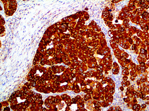
Summary & Explanation
{"type":"root","children":[{"type":"paragraph","children":[{"type":"text","value":"Epithelial Membrane Antigen (EMA) antibody is a mucin-like glycoprotein, shown to be useful as a pan-epithelial marker for detecting early metastatic loci of carcinoma in the bone marrow or liver. It stains normal and neoplastic cells from various tissues, including mammary epithelium, sweat glands and squamous epithelium."}]},{"type":"paragraph","children":[{"type":"text","value":"Hepatocellular Carcinoma, Adrenal Carcinoma and Embryonal Carcinomas are consistently EMA negative, so keratin positivity with negative EMA favors one of these tumors. EMA is frequently positive in meningioma, which can be useful when distinguishing it from other intracranial neoplasms. The absence of EMA can also\n be of value since negative EMA is characteristic of some tumors including Adrenal Carcinoma, Seminomas, Paraganglioma and Hepatoma."}]}]}
Antibody Type
Mouse Monoclonal
Clone
E29
Isotype
IgG2a/K
Localization
Cytoplasmic, Membranous
Dilution
1:250-1:1000
Species Reactivity
Human
Immunogen
Purified human milk fat globule membrane preparation.